Canine Cruciate Disease

Cruciate problems are one of the most common, if not the most common cause of hindlimb lameness in dogs.
The cruciate ligaments in the knee help to provide stability and prevent the thigh bone sliding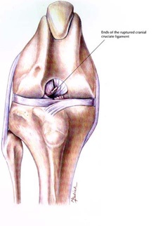 excessively over the surface of the tibia. It is the cranial ligament - the main ligament, that tends to suffer rupture, almost exclusively. In humans, we tend to rupture these ligaments pursuing activities such as skiing, football, etc, but the majority of dogs appear to suffer a gradual degeneration of these ligaments and they can rupture quite early in life.
Diagnosis is often made by clinical examination. We sometimes need to feel the stifle movements with the dog relaxed & this often requires general anaesthesia.
Sometimes, we will obtain joint fluid to support a diagnosis, or in some cases, we may suspect a partial rupture and this does not always leave any laxity, just a rumbling lameness which doesn’t improve. This latter group of patients can be incredibly frustrating to diagnose. Arthroscopy and even MRI scanning are sometimes used !
We have used dozens of techniques over the years. Even human surgeons have an enormous variety of techniques to chose from. We used to use techniques such as wrapping skin, yes skin, through the joint. Injecting it with blood was used. Then we used a technique where tissue from the tissues beneath the skin of the thigh was wrapped through the joint. We have also used nylon, and we still do to mimic the direction of pull of the cruciate ligament. 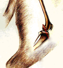
The red line on the diagram roughly shows the location of this.
In recent years, we have turned to techniques which alter the position of the bones, mainly altering the shape of the top of the shin bone, to eliminate movement when the dog stands on the limb. Such techniques have certainly shown a dramatic improvement in the way dogs recover.
The technique I have now turned to has been developed by Dr. Warrick Bruce who happened to have taught me whilst I was at Glasgow University. It is a hybrid technique developed from the research and ideas taken from a number of surgical techniques. It is completed by making three cuts through the tibia, altering the angle of the top of the tibia to try and make this perpendicular to the patellar ligament. Once these cuts are made, the fracture which is created is reduced, i.e. the gap between the bones is reduced and a plate is applied.
The technique has been used extensively by a number of Australian Surgeons with great results and a paper will appear in early 2007 featuring their results.
For a downloadable sheet for prospective patients, click the picture.
I’ll being updating this page with progress at Wright & Morten.
March 2009 - Update
We have now completed some 50 or more procedures with excellent results and no marked complications. Patients have made tremendous progress after surgery and without a shadow of a doubt, their progress is vastly better than that which we used to expect.
Marck 2010
Recently, I completed the official TTA course. More Ts !!
This is the original procedure upon which TTO was based and could be considered slightly quicker and simpler.
The implants are Titanium so they are incredibly strong but their size. They are incredibly thin plates and a cage is used to push the attachment of the patellar ligament forwards. I must say that I really love this technique now but still like TTOs for some patients, and would still choose a different type of surgery for small terriers despite some surgeons finding that this works well.